GENOME STABILITY LAB (GSL)
Research

Genome Stability Lab (GSL)

Research

Research

Research programmes

Our research combines biochemical probe development, proteomics and quantitative microscopy to understand how ubiquitin and ubiquitin‑like signalling controls DNA repair and genome maintenance.
01

How do ubiquitin‑like receptors and transient interactions coordinate DNA repair?

Transient, non‑covalent interactions between ubiquitin‑like proteins and their receptors define recruitment and activity of repair complexes. We aim to map these weak contacts and understand their functional consequences for double‑strand break repair.

Conventional affinity purifications often miss weak or transient binders that nevertheless determine repair outcomes. We use genetic code expansion and site‑selective photo‑crosslinking to capture and stabilise these interactions in cells, followed by affinity enrichment and mass spectrometry to identify bona fide readers.

Identified interactors are validated in cell‑based assays where we quantify recruitment kinetics, influence on end‑resection and impact on repair efficiency using complementary loss‑ and gain‑of‑function approaches. This programme links biochemical mapping to mechanistic cell biology.
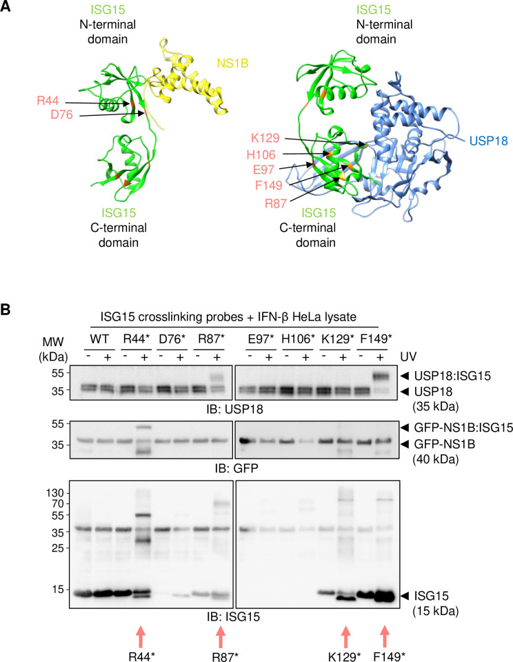
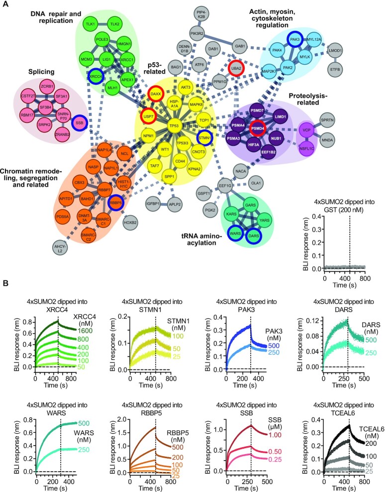
Figure from 'Site‑selective photo‑crosslinking for the characterisation of transient ubiquitin‑like protein‑protein interactions' (PLoS ONE, 2025). Example photo‑crosslinking data and mapped interaction sites illustrating capture of transient, non‑covalent contacts between ubiquitin‑like modifiers and receptor domains — relevant to mapping weak recruitment events at DNA double‑strand breaks.
02

What controls chromatin engagement of non‑homologous end‑joining machinery?

Chromatin context and post‑translational modifications control assembly of non‑homologous end‑joining (NHEJ) complexes. We examine how UBL signalling and chromatin modifiers regulate NHEJ factor engagement at double‑strand breaks.

We combine biochemical fractionation with chromatin‑engagement assays to determine how specific UBLs influence loading and retention of NHEJ factors. Using orthogonal approaches—including proteomics of chromatin fractions and microscopy to follow factor dynamics—we link modification state to repair competence.

Perturbation by genetic or small‑molecule tools allows us to test mechanistic models of recruitment and to identify modifications that alter repair fidelity or pathway preference; findings are tested across cell lines and physiologically relevant cancer models.

Figure 2 from the Nucleic Acids Research (2022) open‑access article describing SUMO‑binding modules and their linkage to NHEJ factor engagement on chromatin (relevant to chromatin engagement of NHEJ machinery).
03

Which E2 enzymes set the stage for repair‑pathway decisions?

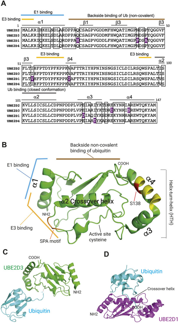
E2 ubiquitin‑conjugating enzymes direct ubiquitylation events that bias repair toward end‑joining or homologous recombination. We dissect E2–E3 axes and their downstream substrates to understand how conjugation specificity impacts repair outcomes.

We perform focused genetic screens and biochemical assays to map functional E2–E3 partnerships. Combining structure‑function mutagenesis with cell‑based reporters for repair outcomes, we track how specific conjugation events alter factor recruitment, end‑resection kinetics and ultimate repair fidelity.

Biophysical and structural approaches complement cellular assays to reveal how E2 specificity and chain topology control recognition by ubiquitin‑binding domains and downstream processing by deubiquitylases.

Figure illustrating structure and functional roles of UBE2D-family E2 enzymes and schematic models of E2–E3–substrate relationships relevant to DSB repair (useful as an open‑access depiction of E2 enzyme mechanisms and context for interrogating which E2s bias repair‑pathway choice). Source: 'The UBE2D ubiquitin conjugating enzymes: Potential regulatory hubs in development, disease and evolution' (PMC9790923).
04

How do deubiquitylating and UBL‑processing proteases sculpt repair complexes?

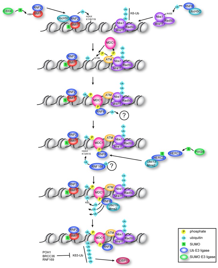
Removal and editing of ubiquitin and UBL signals by dedicated proteases is essential to regulate the lifetime and activity of repair complexes. This programme focuses on proofreading and reversal mechanisms that shape repair dynamics.

We combine targeted protease perturbation with quantitative measurements of factor turnover at damage sites to identify enzymes that regulate signal persistence and repair progression. Proteomic substrate identification is followed by functional testing in cellular assays of genome stability.

Understanding these reversal mechanisms highlights candidate vulnerabilities in cancer where protease activity is dysregulated and suggests routes for therapeutic targeting or biomarkers of repair competence.

Review figure summarising ubiquitin/UBL proteases in DNA repair (open access review figure; see PMC article).